Water Deer - Tusk Construction & Eruption
Table of contents
Canine construction
Teeth are composed of two parts, the root and the crown, the latter section being that which protrudes beyond the gumline. To the best of my knowledge, the minutiae of tusk development has only been studied for a handful of mammals and we don't know what triggers their eruption in most species, including water deer. In dugongs, Elizabeth Burgess and colleagues found that a substantial elevation in testosterone is the trigger, and if canine development is indeed associated with the same pathway as antler formation as Heckeberg postulates, a similar mechanism may be at work in water deer.

Again, as with the spark for their growth, there appears to be very little data on the micrographic structure of water deer canines. We know that most of the crown is composed of dentin, protected by a thin coating of enamel towards the tip, although this can be worn away. In their 2021 paper in the Proceedings of the Royal Society B, Harvard University zoologist Megan Whitney and colleagues note that, owing to the geometric constraints of producing enamel and dentine continuously, the dentine core isn't completely covered with enamel in ever-growing tusks as it is in other teeth. Instead, an enamel cap is present initially at eruption that's eventually worn away, leaving a tooth consisting mainly of dentine with cementum coating the root.
Quite how the tusks are protected from the elements outside the mouth, given that mammal teeth typically require the controlled moisture, temperature and pH conditions within the mouth to prevent drying out and limit dental disease, remains a mystery. Assuming, however, that water deer tusks are structurally in line with those of other mammals, we would anticipate collagen fibrils to form a meshwork between dentinal tubules within the dentin. These fibrils are typically located in two planes, an arrangement different to the single plane set we see in other teeth, and which helps to strengthen the tusk by dissipating inflicted mechanical forces and hampering crack formation. Hokkaido University veterinarian Alireza Nasoori provides an excellent overview of mammalian tusk biology in his 2020 paper in the Archives of Oral Biology.

Nasoori points out that generally a tusk's crown is almost devoid of cells, making cell damage and inflammation less likely in the event of trauma. Furthermore, the dental pulp, the living connective tissue in the centre of the tooth, makes up a proportionally small area that's well separated from the edges. In part, Nasoori explains, these features may minimize the sensitivity of tusks to touch and, more importantly, pain. In elephants, for example, the distal two-thirds of the tusk pulp has very poor innervation, suggesting pain signals are poorly relayed.
In addition to the potential for reduced sensitivity in the tusk itself, the size of the socket (see: Freedom of Movement) and its associated ligament may assist in dampening the force of impacts arising during fights. Finally, the handful of mature skulls I have observed displayed some growth of trabecular bone into the sockets behind the tusks. I assume this is the 'closing of the cavity' that Robert Swinhoe mentioned in 1873, but logically it may also act as an impact-dampening "cushion" of bone, similar to that we see near the base of deer antlers. There doesn't appear to be any special adaptation of the skull itself to accommodate such impact. In 2017, for example, Jinwoo Oh and colleagues studied the skulls of 35 Korean water deer and found no significant difference in the closure rate of the cranial sutures (i.e., the fibrous tissues that unite the bones of the skull and are ossified as an animal ages). The researchers point out that this is interesting because we'd expect the sutures to remain open for longer in bucks than does, to help absorb the forces generated during the rut.

Of course, it may also be the case that the forces are less than we assume, given the tusks are sharp, damage is typically to soft tissues rather than tusks connecting with bone, and the impact isn't directly preceded by a running collision as it is in, for example, sheep.
Tusk eruption and development
The process of tooth formation in mammals is complex and the trigger(s) for their eruption (i.e., the point at which they begin to push through the gum) remains largely unknown. Among the deer, we have a good understanding of the tooth eruption sequence for most species, but their initial development and the catalyst for eruption is, to the best of my knowledge, unknown. Under the assumption that the process follows what we currently understand of the general mammalian scheme, the tusks will develop via a budding of the connective tissue within the mouth, and this probably gets underway in the embryo. They may erupt through the gum via a combination of tusk growth and the shrinking and cross-linking of the collagen fibres in the periodontal ligament (see: Wikipedia - Tooth Eruption for a more detailed explanation of this). This ligamentous connection matters well beyond eruption itself, however: as Whitney and her colleagues note, the mode of attachment for a tusk must be ligamentous (i.e., a gomphosis) to accommodate the sustained movement of the tusk while feeding/chewing throughout life – fusion of the tooth to the jawbone (i.e., ankylosis) would prevent continuous growth.
We do know that water deer tusks grow rapidly from the open hollow root, initially within the large sockets. Indeed, in juvenile animals the root of the tooth is open and straight edged, and the upper tooth walls are thin. (Tusks extracted from juvenile bucks look, to me, more akin to sleeves than teeth.) Data collected on farmland over three consecutive winters, and provided to Arnold Cooke by Cambridge-based deer stalker Martin Guy, suggest the tusks rarely erupt before the bucks are at least five months old. In this dataset, the average length of the canines, from tip to gum line, was only 1 mm (0.04 in.) during November. That said, in his 2019 book Water Deer and Muntjac, Cooke does note that one juvenile buck found dead on farmland in October 1977 had erupted canines, suggesting that they may occasionally erupt before mid-winter. An even more striking case comes from a social media video during July 2026, in which Bedfordshire-based gamekeeper Paul Childerley noted that the tusks were visible in the buck fawn he was hand-rearing at only nine weeks (just over two months) old. From the video, these small white bumps protruded, perhaps, a couple of millimetres below the gum line, though it must be remembered that higher quality nutrition in a hand-reared animal might expedite tusk development over that observed in wild individuals.

Guy's data indicate that following initial eruption, the canine increases in length rapidly, with the mean exposed length reaching approximately 17 mm (0.7 in.) by late December to early January, and around 25 mm (1 in.) by March. Shortly thereafter, typically during April or May when bucks are approximately 10 months old, the tusks begin to protrude below the upper lip and become readily detectable in the field. Nevertheless, I've seen photos of wild individuals from Norfolk, taken in January, that show tusks protruding by approximately 10 mm below the upper lip, despite the bucks being only six or seven months old.
According to Childerley's data from deer shot on the Beckerings Estate in Bedfordshire, this rapid increase in size is initially a reflection of an increase in protrusion, the tusks being similar lengths at the end of November and end of December (around half-grown), followed by a rapid lengthening and increase in curvature. It's worth noting that these are estate-bred animals managed for trophy quality, while Guy's data were from a relatively lightly managed wild population in the Cambridgeshire fens – the two datasets may not be directly comparable as a result. Once the animal is 14-18 months old (e.g., around the end of the following August/September) the base of the tooth begins to fill in, taking on a more rounded shape as the root begins to close. Indeed, writing in 1872, Victor Brooke noted how the canines, as they attain their maximum development, contract their pulp cavity to the point where it is obliterated in old bucks, while at the same time there is a gradual protrusion of the root of the tooth.
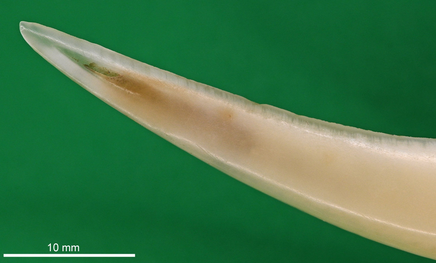
It appears, from the observations of both Arnold Cooke and Paul Childerley, that growth of the tusk after about two years is in the thickness of the root only. Consequently, old bucks may present with chipped and broken tusks, which protrude only a centimetre or so from the gum, but that have very thick roots. Certainly, damaged and even missing tusks are not uncommon in mature animals and, writing to Robert Swinhoe around 1871, Imperial Customs officer Howard Russell confessed that, given how often he found tusks to be damaged or absent, he initially thought they were deciduous until he examined the root of an extracted tusk.
Tusk size
Tusk length appears to be under at least some degree of genetic control, independent of body condition or habitat quality. Game estates that manage bucks for trophy quality are able to produce a disproportionately high number of Gold Medal-standard animals through selective culling – a rate above what unmanaged populations typically seem to produce, and difficult to explain if tusk size were governed by diet and condition alone. Similarly, water deer at Whipsnade Zoo are notably small-bodied, the park being poor habitat for the species, yet long-tusked bucks are not unknown there.
There is considerable variation in tusk length, with 80 mm (3 in.) widely cited in the literature as the maximum exposed length (i.e., from the tip to the gum). Even so, at Whipsnade Zoo, where the overall body size of water deer is comparatively small, Cooke and Farrell (1998) note that tusk length was found to average 44 mm (1.7 in.) during the 1990s, while Cooke's 2019 book Muntjac and Water Deer notes that those in Cambridgeshire farmland averaged 48 mm (1.9 in.), China averaged 53 mm (2.1 in.) and bucks at Woodwalton Fen came in at 60 mm (2.4 in.), the maximum being 72 mm (2.8 in.). Tony Dalby-Welsh of the International Council for Game & Wildlife Conservation's UK Trophy Evaluation Board (CIC UKTEB) told me that the largest tusk measurements recorded in the UK were from a buck shot in Bedfordshire during 2013. The "CIC score" for this Gold Medal buck was 272, made up of lengths of 105 mm/104 mm (around 4.1 in.) and the greatest circumference of each tusk being 32 mm (1.3 in.). These tusks were 4 mm longer than the next highest scoring buck, the circumferences of which were also 32 mm.
The root comprises an additional 1.5 cm (0.6 in.) or so of the aforementioned canine lengths and in mature individuals the cervix, the juncture between the root and crown, is sometimes visible as a dark line running parallel just below the gum. Based on data given by Todd Wheeler in his chapter on sabre-toothed cats in The Other Saber-tooths: Scimitar-tooth Cats of the Western Hemisphere, published in 2011, the periodontal ligament covers about 17 mm of the root – the portion of the tooth embedded below the gumline. Set against the exposed crown lengths described above, that 17 mm represents only around 21% of an 80 mm canine (24% of a 70 mm canine, or 28% of a 60 mm canine). In other words, the ligament-covered root accounts for a comparatively small fraction of the tooth's total length, which suggests that in mature bucks 70–80% of the tusk protrudes above the gumline, though rather less -- perhaps 60–70% -- is actually visible below the top lip, since the lip itself covers part of the upper crown.
When total tusk length is considered -- that is, the measurement taken along the outer curve of the tooth from root to tip -- Cooke and Lynne Farrell, in their account of Hydropotes for the fourth edition of Mammals of the British Isles, reported a range of 81-95 mm (3.2-3.7 in), with a mean length of 87 mm (3.4 in). Variation among mature bucks is well illustrated by two skulls donated to me by Arnold, both from bucks found dead at Woburn during January 1979. One individual, estimated to be approximately 3.5 years old, possessed a left tusk measuring 70 mm in length and 11 mm width at the root, with a mass of 4.3 g. The second buck, estimated at around 4.5 years of age, had a left tusk measuring 81 mm in length and was 15 mm wide at the root, weighing 6.2 g. Thus, the older animal's tusk was approximately 16% longer, 36% wider, and 44% heavier than that of the younger buck.
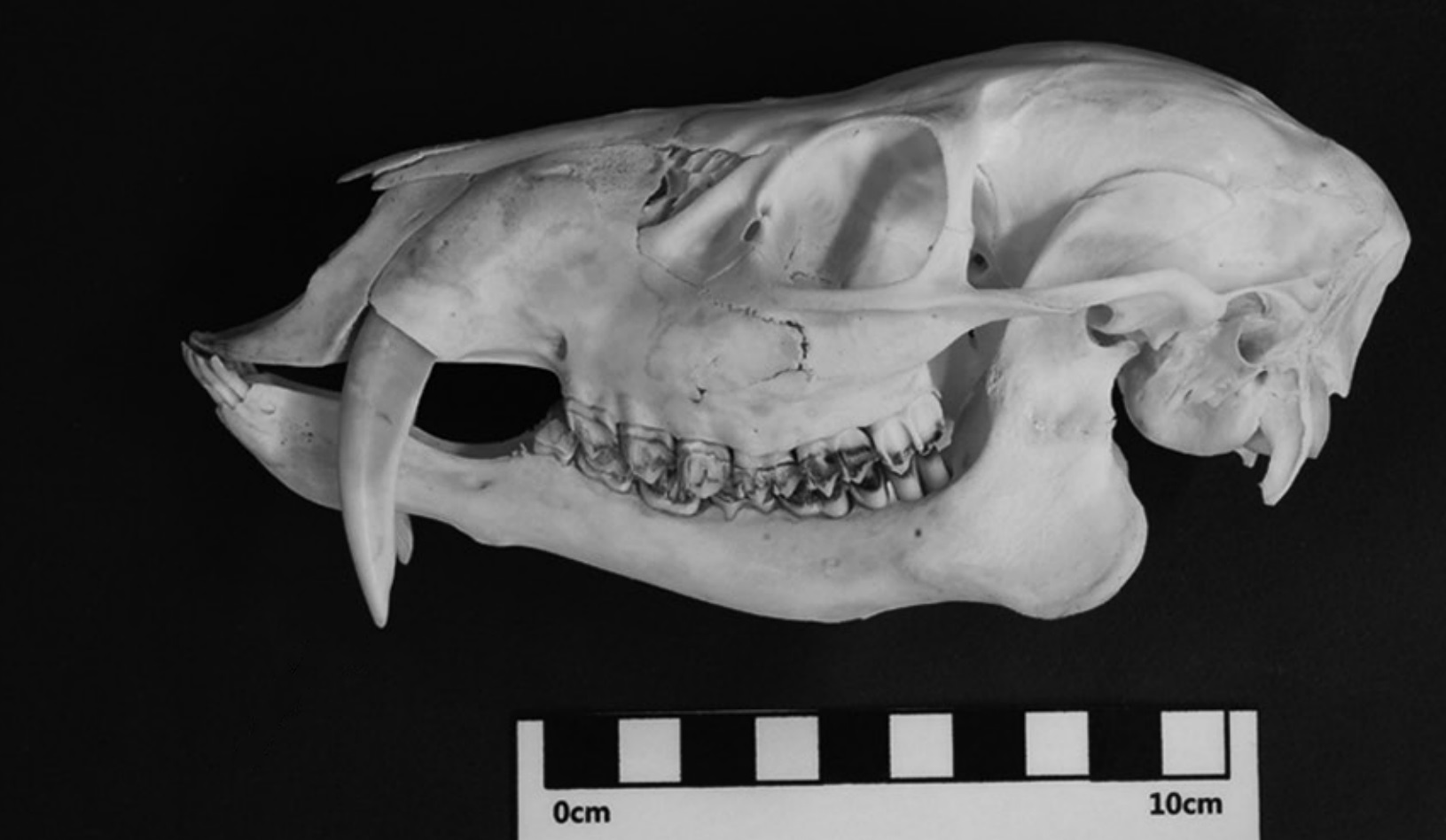
In general, the tusks of mature bucks measure 10-12 mm (0.4-0.5 in) in longitudinal thickness (from front to back), although some individuals occasionally exhibit substantially broader canines. I am aware of at least one animal from Bedfordshire, shot in February 2021, that had tusks of average length but measuring 16 mm (0.6 in) in width. Similarly, in a 2013 paper published in the Journal of Veterinary Medical Science, Yungkun Kim and colleagues included a photograph of the skull of a Korean water deer buck with a comparably thick canine (above). In such cases, the inner edge of the tusk is noticeably straighter than that of narrower examples. I have no equivalent tusk size data from Norfolk, Suffolk, or Buckinghamshire and would be interested to hear from anyone who does.
According to the requirements set out by the CIC UKTEB, to qualify for the lowest level of "medal" (i.e., bronze) a buck requires a canine length of at least 65 mm (2.6 in.) and a circumference of at least 25 mm measured at the point along the tooth where its diameter -- viewed side-on -- is greatest.
References
Burgess, E.A. et al. (2012). Testosterone and tusks: Maturation and seasonal reproductive patterns of live, free-ranging male dugongs (Dugong dugon) in a subtropical population. Reproduction. 143(5): 683-697.
Cooke, A. (2019). Muntjac and Water Deer: Natural history, environmental impact and management. Pelagic Publishing, Exeter.
Cooke, A. & Farrell, L. (1998). Chinese Water Deer. The Mammal Society & British Deer Society.
Cooke, A. & Farrell, L. (2008). Genus Hydropotes. pp. 617-622. In: Harris, S. & Yalden, D.W. (eds.). Mammals of the British Isles: Handbook, 4th Edition. The Mammal Society, Southampton. 800 pp.
Kim, Y. et al. (2013). Skull growth of the Korean water deer, Hydropotes inermis argyropus. Journal of Veterinary Medical Science. 75(7): 867-878.
Nasoori, A. (2020). Tusks, the extra-oral teeth. Archives of Oral Biology. 117: 104835.
Oh, J. et al. (2017). Cranial suture closure patterns in water deer and implications of suture evolution in cervids. Mammalian Biology. 86: 17-20.
Swinhoe, R. (1872). Notes on Chinese Mammalia observed near Ningpo. Proceeding of the Zoological Society of London. 40(1): 813-818.
Swinhoe, R. (1873). On Chinese Deer, with the description of an apparently new species. Proceeding of the Zoological Society of London. 41(1): 572-576.
Wheeler, H.T. (2011). Experimental paleontology of the Scimitar-tooth and Dirk-tooth killing bites. p. 19-34 in Naples, V.L., Martin, L.D. & Babiarz, J.P. (eds). The Other Saber-tooths: Scimitar-tooth Cats of the Western Hemisphere. Johns Hopkins University Press, Baltimore. 252 pp.
Whitney, M.R. et al. (2021). The evolution of the synapsid tusk: insights from dicynodont therapsid tusk histology. Proceedings of the Royal Society. 288B(1961): 20211670.