Water Deer - Tusk Movement
While any large teeth are beneficial in the right circumstances, they can also be an impediment. One way these teeth can reach their full potential is if they're mobile. If the tusks aren't fixed to the jaw they're flexible to move back and forth and side to side, allowing them to move out of the way while feeding and, of particular importance for a ruminant that spends a lot of its time chewing cud, they don't get in the way of sideways jaw movements. It is just such mobility that we see in the tusks of mature Chinese water deer, although the mechanics of it remains a matter of debate. It is worth noting, at this point, that only fully developed tusks (i.e., completely erupted canines with closed roots) are mobile, immature tusks are not.

The sockets in the skull in which the tusks sit are referred to as maxillary alveola, basically 'air sacks' within the maxilla (upper jaw), and those of water deer are exceptionally long and wide. The upper reaches of this are often visible as a mild bulge in the skull of the male and, in the chapter on artiodactyl teeth in their The Teeth of Mammalian Vertebrates, Barry Berkovitz and Peter Shellis include a radiograph of the skull of a mature buck highlighting just how large the alveola are; their rear wall extend back to the juncture between the second and third premolar. There's no scale on the radiograph but, based on the tusks and other cranial proportions, I would estimate that each alveola was approximately 50 mm (2 in.) long and 20mm (0.8 in.) at the widest, which corresponds with the handful of skulls I have seen.
Despite having a short root and being remarkably mobile, they are surprisingly difficult to extract. Indeed, former Zoological Society of London veterinary officer James Aitchison, writing in 1946, recounted how, on one occasion, a water deer at Whipsnade tried to climb a strong wire-mesh fence and ended up hanging by its tusks on one of the wires; testament, if any were needed, to how strongly embedded the tusks are. In his 1973 book, Beasts in My Belfry, Gerard Durrell recounts what appears to be the fence-hanging story, which began with him and three colleagues trying to net a buck who had escaped the park's perimeter fence and managed to get into a chicken run in a nearby field during the winter of 1945/46:
"The Chinese water deer became more and more panic-stricken and started hurling himself at the tall wire fence in an effort to break through. He made one prodigious leap, hit the fence and hooked himself neatly on to the mesh by his two tusks and hung there kicking and struggling. We made a concerted rush but at the last minute he managed to unhook himself with a magnificent display of muscular contraction and, landing on the grass, turned with tremendous speed and broke through out ranks."

The reason behind the security of the tusk is a very thick pericementum, the collagen-rich fibrous connective tissue otherwise known as the periodontal membrane or periodontal ligament surrounding the tooth. This ligament holds the tooth extremely securely and is itself firmly attached to the walls and outside edge of the socket. The only measurements for the periodontal ligaments surrounding the root inside the sockets that I have come across in the literature to date were given for a mature buck by Todd Wheeler, a palaeontologist at the Powell Museum in Los Angeles, in his chapter on the killing bites of sabre-toothed cats that was published in 2011 as part of The Other Saber-tooths: Scimitar-tooth Cats of the Western Hemisphere. Wheeler reported the ligament to be just over 3 mm (0.1 in.) thick and nearly 17 mm (0.7 in.) wide, covering virtually the entire root. This aligns with my observations of tusks I have extracted from road-killed animals.
While the alveolar capacity and periodontal ligament permit some mobility, it's rather limited and there is a 'hard stop' as the root connects with the walls of the socket. One of the earliest and most detailed studies on the tusks of deer was carried out by Zoological Society of London vet James Aitchison based on two fresh heads, one skull, and "several live" water deer at the Society's Whipsnade Zoo. Aitchison published his findings in a paper to the Proceeding of the Zoological Society of London in 1946, in which he noted that the slightest pressure moves the tusks by 15 mm (0.6 in.) front-to-back and 25 mm (1 in.) side-to-side, corresponding with my observations and those made by Barry Berkovitz and Peter Shellis in 2018. Aitchison observed that when the pressure was removed, the tusk didn't return immediately to any fixed position, but moved very slowly towards the centre of the ellipse of movement. In other words, it didn't spring back to position as might be expected if the stretch of some highly elastic ligament were responsible for the movement.
An area of some contention among observers has been how much of the tusks' movement is under the control of the buck. In the late 1800s, Swinhoe noted how, during his observations of a captive animal, he never once saw it move its tusks, which always appeared to be held back. In dead animals, by contrast, Swinhoe had observed the tusks were always vertical, suggesting the "pulling back" was an active engagement by the buck. As such, he sent a fresh head of a buck to anatomist Dr. R. Jamieson in Shanghai who dissected the tusk and examined the surrounding tissue under a microscope. Jamieson's findings failed to support Swinhoe's hypothesis, concluding:
"... the movement of the tooth moves the surrounding gum, which firmly clasps it; but neither the gums nor lips have any power to move the teeth."
Some seven decades later, Aitchison described how the gum terminates along its lower border, next to the gum pad, in a thickened band that formed a loop, also freely movable, around the distal surface of the tusk. In other words, this tough band forms a "lasso" around the neck of the tusk and, in his paper, Aitchison proposed:
"When the animal snarls, the snarling muscles raise the upper lip and pull the moveable gum forwards and upwards. The thickened band and loop of the gum, in turn, hinge the tusk forwards into its most 'erect' position before the animal 'strikes'."
The handful of examples of bucks fighting of which I'm familiar support Aitchison's description and writing in their Mammals of the British Isles: Handbook, 4th Edition chapter, Arnold Cooke and Lynne Farrell described the same scenario:
"This snarling action pulls forward a section of movable gum which brings the tusk into its most forward position."
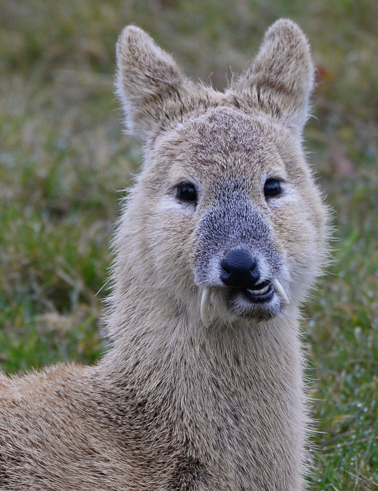
In discussing this with Dr Cooke, he pointed out how the snarling action results in a change in the profile of the nose and, at the same time, a parting of the lips and rotation of the ears to point forwards. Indeed, in the superb series of images captured by Mike McKenzie at Claxton in south-east Norfolk during February 2013, the shortening of the nose and pulling back of the lower jaw, slightly parting the lips, can be clearly seen. This has the effect of bringing the tusks into a near-vertical position and actually gives the illusion of more movement in the tusks than the anatomy would seem to allow.

Based on Aitchison's findings, once the buck relaxes, the thickened gum pad (i.e., the periodontal ligament) then slowly returns the tusk to its resting position which, again going by Aitchison's observations, is around the middle of its full range of movement. This corresponds with video footage of freshly shot bucks Arnold and I have seen. Some observers suggest that the tusks are actively "pulled back" out of the way while grazing, but to the best of my knowledge no mechanism has ever been documented to support this. Indeed, Arnold's and my observations indicate that the relaxed/grazing position may be at or just behind the middle of the orbit, and any movement further backwards might simply be a reflexive response to mouth movements associated with grazing, rather than being a conscious effort on the part of the buck. During our discussion on the topic, Arnold suggested to me:
"Maybe the animal cannot control further backward movement, but such involuntary movement exists to reduce the risk of the tusk breaking when fighting or simply going about its usual business. We know tusk breakage happens fairly frequently, so a mechanism that reduces the risk must be a good thing. In other words, the grazing position is the central usual position; the animal can bring the tusks forward by snarling; the buck cannot make them go further back from the central position, but if a tusk is knocked or if someone pushes it, such movement is possible."
Subsequently, Arnold went on to muse about whether the stretching down of the neck while the buck is feeding might affect tusk position. Conceivably, he postulated, stretching the neck might "shift things around in the jaw of the deer so the tusk goes back".

My cursory analysis of video stills and photographic images indicates that the posterior edge of the tusk emerges from the upper lip at an angle of approximately 40° in grazing males, around 50° in relaxed individuals (e.g., resting, walking, or recently shot), and up to approximately 70° when protruded during a snarl. Although tusk length and curvature vary among individuals, this preliminary assessment suggests that the tusks are positioned slightly further posteriorly during grazing than in a relaxed state, with the relaxed position lying near the midpoint of their apparent range of movement.
It remains unclear whether this more posterior position during grazing reflects active muscular control by the buck, or is instead a passive consequence of head posture and associated muscular or ligamentous tension during feeding. At present, there is little evidence to support the existence of fine voluntary motor control over tusk position. Indeed, observations reported by Swinhoe and, more recently, by Todd Wheeler, a palaeontologist at the Page Museum in Los Angeles, indicate that the tusks are not independently movable. Rather, anterior projection appears to occur only in association with snarling, with a return to the resting position following relaxation; movement of the tusks between these positions does not appear possible in the absence of these behaviours. Dr Wheeler told me:
"I couldn't find, and can't imagine, any provision for voluntary movement within the periodontal ligament."

In addition to the limited anterior displacement of the tusks prior to combat, anatomical evidence suggests that relatively weak masseter (chewing) musculature may facilitate their effective use by permitting a greater degree of jaw opening. In a 2013 study of the water deer masseter muscle, Motoki Sasaki of Obihiro University of Veterinary Medicine and colleagues reported a weakly developed maxillo-mandibularis tendon -- the structure anchoring the masseter muscle to the lower jaw -- which they proposed may allow a wider-than-typical gape. If this interpretation is correct, such an adaptation could enhance the functional deployment of the tusks during combat, in a manner comparable to that observed in Bactrian camels (Camelus bactrianus). Behavioural observations further support this possibility: on two occasions I have witnessed bucks opening their mouth widely when facing an opponent during fights, suggesting that gape may form part of the threat display and/or facilitate effective tusk contact. Unfortunately, the anatomical study by Sasaki and his team was based on only two female individuals recovered in Gyeonggi-do, so we don't know if the same applies to bucks.