Part 2: The smell of fox: Types of scent produced by foxes
It's difficult to discuss the scents and pheromones produced by foxes without delving into the chemistry, particularly as many of the volatiles we know they produce have no pithy or colloquial abbreviations. I list some of the chemicals here simply for completeness, but I have made few attempts to discuss them in any great detail. For the purposes of this article it is sufficient to recognise that these compounds are present, and the more chemically-minded reader is directed either to the primary literature referenced in the main text, or to contact me for my research notes for this Q&A which contains more details and a bibliography.

Much of what we know about the chemical composition of the urine and faeces of the red fox comes courtesy of the unenviable studies Eric Albone, James Jorgenson and the late Jackson Laboratory biologist Wesley Whitten carried out during the 1970s. Similarly, we owe a great deal of our understanding of the constituents of the anal sacs to the work of Georges Ware and Pauline Gosden at Bristol University, and that of the violet gland to University of Saskatchewan biologist Peter Flood and German anatomists Peter Meyer and Helmut Wilkens. It is on the work of these scientists that much of the below is based.
Finally, it's important to recognise that, while we have an increasingly clear picture of the volatile chemicals produced by foxes, our understanding of how foxes interpret these messages - i.e., what these different chemicals tell one fox about another - is still very much in its infancy. Similarly, it seems likely there are still more components left to identify in scent marks. The work of the aforementioned pioneers during the 70s documented a dozen or so constituents in urine and faeces, while more recent data collected by Eleanor Kean at Cardiff University suggest fox scent is a much more complex mixture of chemicals than first thought - her initial GC-MS scans of swabs taken from fox cheek, anus glands and faecal samples identified at least 80 different compounds. Kean's preliminary findings were presented as part of the Foxes Live series that aired on Channel 4 in the spring of 2012 but unfortunately the programme makers only funded the initial analysis, and no further funding has been available to complete and publish the work.
Urine and faeces
The strong “skunky” or “musty” smell in a damp woodland. The pungent dark droppings that grace visually conspicuous or unusual objects in the landscape and that your dog makes a beeline for. Most of us are familiar with this aromatic collateral of having foxes in our neighbourhood. For the “vulpiphiles” among us, these smells are handy as they allow us to track foxes, establish their routes and map out their territory configuration. For others, this affront to our senses is just another reason to lament having them visit. Indeed, in a recent survey of households in south-east England conducted by Surrey-based ecologist Adele Brand and myself, 20% of respondents who provided reasons for their dislike of foxes cited fox poo.
Writing in the journal Science in 1978, Indiana University chemist James Jorgenson and his colleagues described their analysis of fox urine samples collected from the snow in and around Acadia National Park, Maine. They identified that the pungent, skunk-like quality of fox urine is primarily a result of two chemicals, Δ3-isopentenyl methyl sulfide and 2-phenylthyl methyl sulfide, both of which generate a strong sulphurous odour.

Other compounds isolated from the urine by Jorgenson's team, and subsequently Whitten and his colleagues in a paper to the Journal of Chemical Ecology in 1980, include 4-heptanone (aka butyrone, a fruity-smelling compound), 6-methyl-5-hepten-2-one (known as sulcatone, also component of human sweat with a vaguely citrussy odour), benzaldehyde (almond-scented), 1-phenylethanone (floral-scented acetophenone), 2-methylquinoline (quinaldine, which also occurs in tea plants) and trans-geranylacetone, which has a floral-fruity odour.
Whitten and his co-workers suggested that one particular chemical, quinaldine, may be sex-specific having only found it in the urine of dog foxes. An ensuing study of the urine of four captive fox cubs by a team of biologists, led by Sidney Bailey at the Ministry of Agriculture Fisheries & Food in 1979, however, also found quinaldine in females, although it should be noted that the content of urine appears to vary seasonally in both sexes. Indeed, Bailey found 4-heptanone increased in concentration from December to a peak in mid-February (the breeding season) and remained relatively high until April. Whitten and colleagues observed 3-methylbutyl methyl sulfide for the first time in the urine of both sexes and found it peaked during February in males.
When it comes to the chemical composition of fox droppings, fewer studies have been conducted, and things get a little more complicated because the faeces themselves may be augmented by liquid ejected from paired anal glands, situated either side of the anus (see below). Hence, chemicals isolated from scat may not necessarily have been released in them but squirted on them afterwards.

In 1980, Evelyne Vernet-Maury at the Université Claude Bernard in Lyon reported 2,4,5-trimethylthiazoline (TMT), a substance with a nutty/chocolaty smell to it, in fox droppings and subsequently suggested it was this component that caused a fear response in the brown rats in her lab. Subsequently, in a paper published in 1984, Vernet-Maury and two colleagues noted that some of their unpublished research found around 70 volatile components in fox faeces, including 1-methylmercapto-3-methylbut-3-ene and 4,4-dimethyl-1,2,3-trithiane. 3,3-Dimethyl-1-2 dithiolane, a substance found in coffee, and 4-Mercapto-4-methylpentan-2-one, which the authors described as having an “odor reminiscent of mature tom-cat urine and of heated asphalt tar” were also recorded. Analysis of 24 samples of fox poo collected from animals in another facility in France by Olivier Rampin and colleagues Université de Paris-Saclay, however, failed to find TMT, although they do concede this may reflect their GC-MS method being insufficiently sensitive. Unfortunately, Rampin don't list any of the substances they did find in their 2018 paper to Chemical Senses.
Kean, in her Foxes Live summary, reported having isolated 80 compounds in the scent samples she analysed including indole, acetone, phenol and benzaldehyde, but didn't specify which compounds were extracted from which samples. Indole gives mammal faeces the characteristic poo smell, so it seems likely to be present in fox droppings.
Anal glands
Beneath the skin on either side of the anus of a fox, between the inner and outer anal sphincters, sits a roughly spherical gland, approximately 10mm (0.4 in.) in diameter, that holds up to about 1 mL of pungent-smelling watery yellowish fluid; a “bitter-smelling milky fluid”, as Lucy Jones called it in her book, Foxes Unearthed. These are the anal glands, or anal sacs. The glands empty into the inner cutaneous anal zone via a short duct. Based on Berlin University anatomist Ingrid Spannhof's examination of 14 (8 female, 6 male) foxes collected from German zoos and David Macdonald's excision of 29 (17g, 12m) wild foxes killed in Oxfordshire the walls of the anal sacs are richly supplied with apocrine (secretory) glands whose ducts empty into the lumen of the sac with epithelial cells noticeably fatter and with larger nuclei during the winter breeding season. The striated muscle interspersed amongst the tubules presumably serves to evacuate the sac's contents.

We assume that the anal sac secretion is either imparted on faeces as it moves through the anal canal or deposited on them afterwards. As far as I know, however, this has never been empirically studied in foxes as it has for big cats. Using an inert green food dye, Cheryl Asa at the St Louis Zoological Park in Missouri found that no anal sac secretions were detectable in any urinary or faecal markers of cheetahs, tigers or leopards, suggesting the anal sacs are expelled onto the droppings after they've left the body. Indeed, Eric Albone suggests that about 0.5 mL of this liquid is often, but not always, squirted onto faeces, and in her book, The Nature of Foxes, Rebecca Grambo notes that it appears more commonly deployed by males than females. It is presumed that this fluid adds to the message encoded in the droppings.
Work by Albone and his colleagues has given us good insight into the conditions inside the sacs and the composition of the fluid they produce. The sac environment and secretion is alkaline (pH 6.5-9.4), which is important because acidity affects the so-called “free acid composition” that ultimately determines the odour of the substance. The glands themselves are home to a community of microorganisms that appear to generate the specific odour contingents of the secretion.
In their chapter in Flavor Chemistry of Animal Foods, published in 1978, Albone and his co-workers described the fox anal sac as a highly reducing environment (redox potentials down to -400mV), suggesting that the microorganisms present, specifically streptococci and Proteus, create and maintain anaerobic conditions (i.e., low or no oxygen present) in which strict anaerobes can thrive. These “obligate anaerobes” tend to be very active odour producers. The microbiologists noted that the anaerobic microflora largely comprised Clostridium and Eubacterium, with Bifidobacterium, Fusobacterium, Bacteroides and Peptostreptococcus sometimes present in lower concentrations. A subsequent study by Georges Ware and Pauline Godsen at Bristol University found these same six bacteria, with Clostridium perfringens the most commonly observed species, and reported vast numbers of these obligate anaerobes in the fluid - billions per mL.
The anal sacs do contain some aerobic microflora, the community dominated by streptococci (mostly Enterococcus (formerly Streptococcus) faecalis) and Proteus (mostly Proteus mirabilis). Curiously, however, coliforms and gut lactocacilli are largely absent, despite the sac being close enough to the anus to permit passage of faecal flora into it. Inoculation studies by Albone and his colleagues suggest that the reducing nature of the anal sac makes for a highly selective environment for bacteria. When the team injected a solution containing eight billion Escherichia coli per mL into a sac, they found their population had reduced to only 4% after four days, and E. coli was absent two weeks later. Over the same period, populations of Enterococcus/Streptococcus and Proteus bacteria remained stable. Conversely, E. coli are abundant in fox droppings, while Enterococcus/Streptococcus and Proteus are rarely found, suggesting a different microbial environment in the anus itself.
The gland secretion is composed primarily of volatile fatty acids (C2-C6, particularly acetic, propionic, isobutyric and valeric acids), which are produced when microbes breakdown proteins. We know that diamines (putrescine [1,4-diaminobutane] and cadaverine [1,5-diaminopentane]), indole and ammonia are also present, which we think are probably produced by the microbes too. Some studies have found trimethylamine, soluble protein and 5-aminovaleric acid, the latter of which seems to play a role in regulating bacterial species composition. Additionally, Eric Albone and Truls Grönneberg observed levels of free and mean cholesterol (56% and 0.93 mg/g, respectively) much lower than that found in domestic dog anal sacs (66% and 2.49 mg/g). The secretion itself also contains variable quantities of cell debris and lipids, known or suspected to be produced by anaerobic microflora.
We know that these volatile components of the gland secretion are produced by the microbial community in the sac because when Albone and his colleagues sterilized the sacs of some captive animals it took at least three days for fatty acid production to resume as the microbial assemblage recovered.
The composition of the secretions from the right and left gland usually similar, although not invariably so. Eric Albone and George Perry found, for example, twice as much cadaverine in the right sac as the left in some of their subjects.
The violet (supracaudal) gland
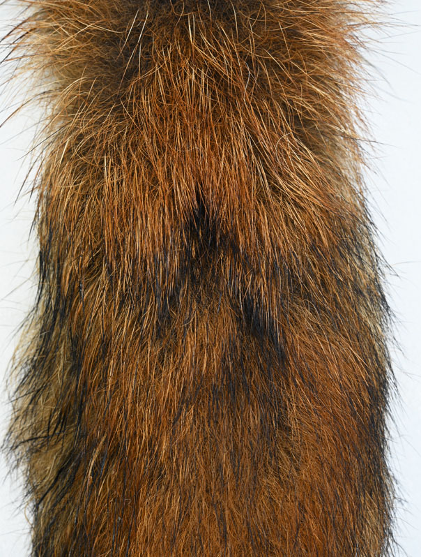
A large gland is present on the upper surface of the tail in both sexes. In the literature this gland is variously referred to as the supracaudal gland, violet gland or simply “tail gland” and the earliest description I can find was written by German theologian Caspar Schwenckfeld in his Therio-trophevm Silesiæ, a Latin tome published in 1603 on the “trophy beasts of Poland”, which included the red fox. Loosely translated, Schwenckfeld wrote of the fox that:
'Nature has given him a tail, and a tuft of thick, yellow hair grows in the higher slender part, called 'the flower'; its March violets smell deceives the hunters of our country.'
Seventy-four years after Schwenckfeld referenced this “violet gland”, Danish anatomist Caspar Bartholinus considered the gland to smell of violets and described bristle-like hairs sticking out of it. Huw Lloyd in his 1980 book Red Fox references “Hartig (c. 1830)” calling the gland Der Voile after its distinctive flowery smell. (I presume Lloyd was referring to German forestry biologist Theodor Hartig, but he does not cite the source in his book and I have been unable to track down an original reference for this.) Lloyd himself wrote:
“Although I have investigated the region of this gland in dead foxes with my nose, I have been unable to detect anything unusual, but colleagues persuaded to investigate likewise have described the smell as not unpleasant, without being more specific.”
Several other early naturalists verified Bartholinus' observations that the gland was populated with hairs. Writing in 1848, Swedish biologist Anders Retzius described colourless hairs with black tips in the area of this oval gland, devoid of underfur, which measured 26 x 9mm (1 x 0.35 in.). Retzius considered, however, that the gland had a smell more similar to rock lichen than violet. Five decades later, in 1899, British zoologists William Edward de Winton, wrote of straight coarse hair in the area of the gland with a nearly white body and black tip.
German anatomist Karl Toldt, at the Imperial Natural History Museum in Vienna, provided some additional information on the supracaudal gland in 1937, describing a spot on the tail about 15mm (0.6 in.) in diameter situated on the upper side of the tail, about 6cm (2.4 in.) behind the root, and consisting of a cluster of large and small sebaceous glands apparently producing a fatty substance of unknown function. The hairs in the region of this “lancet-shaped” organ were taut and colourless except for the black tip - the hair body is bright, translucent and yellowed by the glandular secretion, which may form irregular yellowish granules on and between the hair, according to Toldt.
Retzius considered that dog foxes possessed a larger violet gland than vixens, but Toldt failed to find any variation conclusively linked with sex in the 190 glands he studied, although he did note that there was considerable variation in size and shape among individuals.
A more detailed description of the tail gland was presented by Josef Schaffer in his extensive review of mammalian skin glands, published posthumously in 1940. In the review, published in German, Schaffer described extensive glandular bodies, pigment-less hairs and powerful bundles of muscles. The individual glandular lobes showed differences in structure and secretion type with both season and sex, and he found a “greasy” secretion in the vixen and “protein-like” secretion in the dog. Given the difference in seasons over which his samples were collected, Schaffer concluded that the glands are transformed to produce fatty secretions in winter.

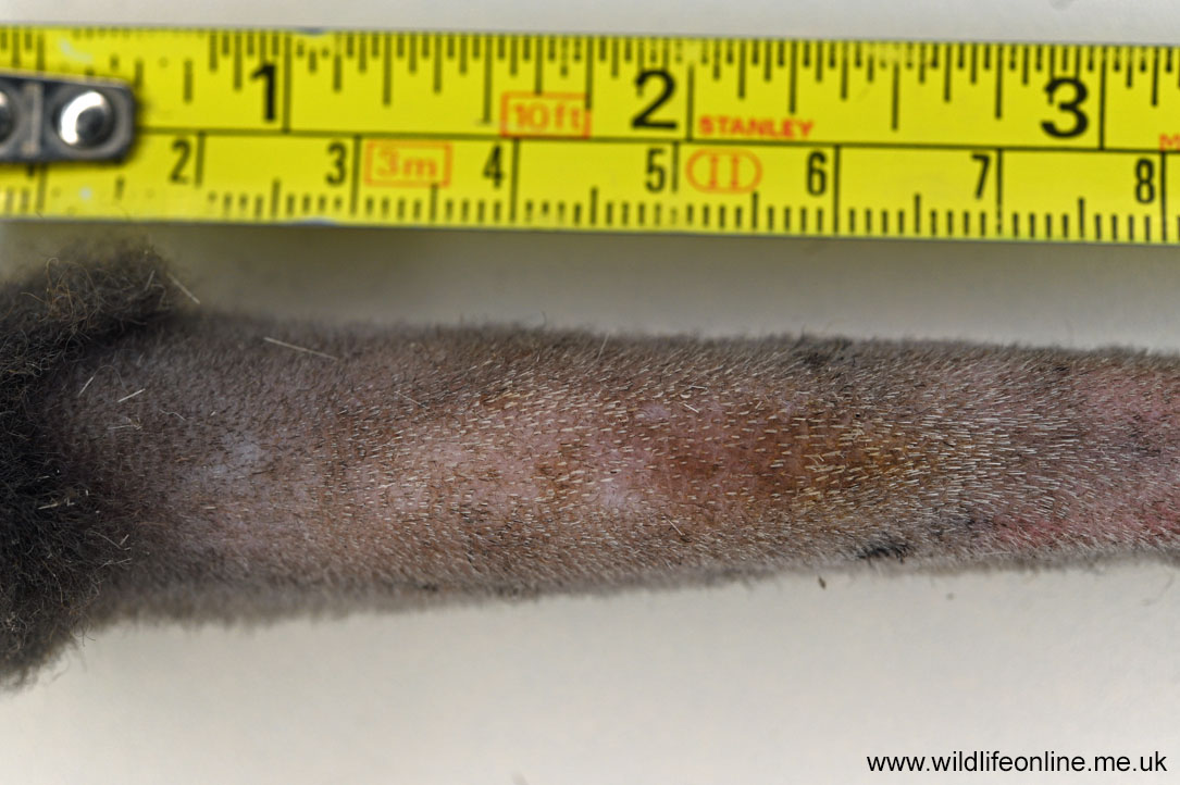
The earliest attempt to comprehensively study the gland microscopically and histologically of which I am aware was conducted by German anatomists Peter Meyer and Helmut Wilkens in 1971. Their study, also in German, documented this “viola” located 10-11cm (4 in.) behind the tail root, over the seventh tail vertebrae, visible in the fur as a black spot (the “violfleck”) 2-5cm long by 1-3cm (1.5 x 0.8 in.) wide. In most foxes, even as young as three months old, the gland was very noticeable, while it was faint in two of their adult subjects and, because of the way the coat flows, the spot is about 3cm behind the gland itself. In common with earlier observers, Meyer and Wilkens noted a dense bundle of bristle-like guard hairs embedded in the gland and a lack of underfur. The guard hairs had pigmentless shafts except for a black tip, were 6-7cm (2.7 in.) long, causing them to protrude some 3-5cm (2 in.) beyond the rest of the coat, and 90-150µm wide (i.e., if the widest ones were lined up next to each side by side other you'd have 170 in an inch).
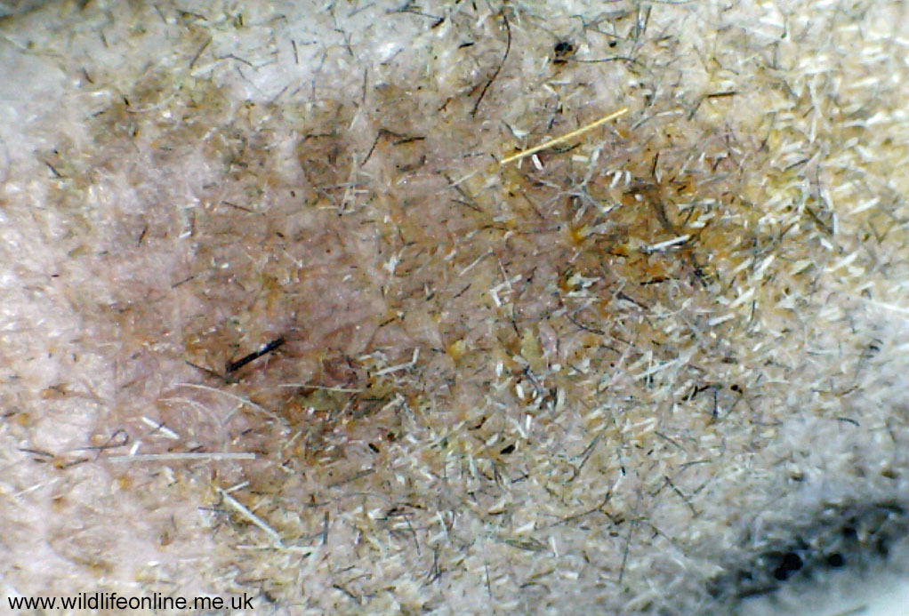
Upon parting the violfleck fur, Meyer and Wilkens recorded an oval/elliptical-shaped gland in the skin visible to the naked eye - pinkish in colour, 22-30mm long, 6-10mm wide (1.2 x 0.4 in.) and about 3mm deep. The skin around the gland swells according to the season and can be partly obscured by the greasy yellowish lipoprotein substance that it secretes. In the summer Meyer and Wilkens barely observed any secretion, while in the winter it would sometimes form a layer on the skin up to a millimetre thick. The substance itself is squeezed out onto the skin by the unusually powerful arrector muscles surrounding the gland, and sticks to both the skin and guard hairs. It is massaged up the shaft of the hair by their reciprocal movements, helped along by their highly sculpted surface.

The hairs have split tips, which we think promotes adhesion and dispersion of the secretion and its scent. The secretion seems to turn dark upon exposure with air or light, and Meyer and Wilkens reported that the guard hairs often developed a fine-skinned black-grey coat extending almost from the tip down to about 1cm above the skin. As the secretion dries on the skin it flakes, leaving yellow-brown waxy granules that retain their odour. Presumably this waxy sebum is the reason the woolly underfur is absent around the gland; it would get gunged up.
The gland itself is composed of a few tubular apocrine sweat glands that are buried deep in the skin, the ducts of which pass through an abundance of massive multi-lobed sebaceous glands and open into the hair follicles. The gland has a unique steroid metabolism, and chemical analysis of the waxy secretion by Eric Albone and Peter Flood found it to contain several volatile terpenoids, breakdown products of carotenoids that chemists collectively refer to as apocarotenoids and that are often associated with fragrances. In particular, dihydroactinidiolide (DHA) and trans-4-keto-β-ionone were identified. These apocarotenoids are also found in plants; the ionone structure gives flowers of violets (Viola spp.) their characteristic smell and presumably explains the scent described by early authors. The gland was also found to have hydroxysteroid dehydrogenase activity (a particularly strong β -3 β HSD reaction for the chemists among you), suggesting that it is related to sex hormone production and/or metabolism, and to be rich in glycogen.
We know that animals can't produce carotenoids and must get them through their diet. As such, Stuart McLean and his colleagues, writing in 2019, suggested that, as foxes eat a significant amount of fruits and seeds, they likely have a diet supplying common carotenoids. Mammals are well-known to have the biochemical machinery to cleave carotenoids into apocarotenoids, after which they can be modified to allow skin cells to take them up, and McLean and his team proposed:
“… that these processes produce the floral scents of the fox tail gland, rather than foxes, alone amongst animals, synthesize apocarotenoids directly.”
Assessing various skin glands of four foxes shot as part of a culling plan on Australia's Anderson Peninsula in southern Victoria, McLean, at the University of Tasmania, and colleagues observed that all the redolent compounds they found (broadly terpenes, monoesters and diesters) were more abundant in the tail gland than on the flanks or muzzle. A total of 98 fatty acids were found; 85 were present on the tail gland fur, 26 of which were only found on the tail gland. Branched fatty acids were most abundant on the tail gland fur, while unbranched acids were more prevalent on the flanks and muzzle. Terpenes, aromatic organic hydrocarbons often encountered in essential oils, were also more abundant around the tail gland than at either other site.
Finally, a curious feature of the supracaudal gland is that it appears to glow under ultraviolet light. Albone and Flood, in their 1976 paper to the Journal of Chemical Ecology, noted a strong yellow-green fluorescence of viola tissue under UV light, and that the tissue of males fluoresced more than that of vixens. The cause of this “glow” is unknown, although Albone and Flood suggest it may be a result of β-carotene, or at least carotenoid in origin. Whatever the cause, the authors considered it unlikely that this fluorescence was visible to the foxes and more likely a by-product of the gland's operation. Coincidentally, I have noticed that this area of fur is clearly visible as a black spot on infrared trailcam footage (suggesting it absorbs light at around 940nm) and I have seen thermal images in which the tail gland has shown up as significantly warmer than the surrounding coat, presumably because the secretion remains warm for a while after excretion and reflecting the lack of underfur making that section of the tail less well insulated.
Other glands

In his Russian book Mammal Skin, published in 1982, Vladimir Sokolov described small (up to 30µm in diameter) and sparsely distributed sebaceous glands on the body, and very large (up to 200µm) multi-lobed glands concentrated on the soles of the feet. In Running with the Fox, David Macdonald noted that fox feet have deep interdigital (i.e., between the toes) cavities in which the highly glandular skin is tinged red. In his 1933 book Hunting by Scent, former Master of the Bicester and Warden Hounds Hubert Budgett described a “scent oil” secreted high up between the toes, close to where they join the pad, and impregnating all the hairs covering the foot. It is this scent trail that hounds follow, although, on his slides, oil from the pads dried within 3.5 hours, suggesting the trail can “go cold” relatively quickly. In addition to this, Huw Lloyd, in his 1980 Red Fox, noted that the heavily cornified skin of the foot pads contains true merocrine sweat glands, although he did not know whether these were associated with scent marking.
Both Sokolov and Macdonald refer to a subcaudal (“under tail”) gland, similar in structure to the violet gland but composed largely of big sebaceous glands with large tubular apocrine glands underneath; apparently, it's similar in structure to that found in goats. Sokolov also noted that the state of the skin changes throughout the year, based around three main “phases”.
| Period | State of Skin |
|---|---|
| Late November to February | The skin, dermis and epidermis are all thin, the stratum corneum well developed, sebaceous glands large and very active while apocrine (sweat) glands are only slightly developed. Arrectores pilorum muscles that control the hairs are powerful. |
| March to June | Foxes undergo partial moult and sebaceous glands atrophy while sweat glands increase in size. Dermis and stratum malpighii of epidermis thicken. Arrectores pilorum becomes thin, as does subcutaneous tissue layer. |
| August to early November | Full moult occurs and all old fur is replaced; new fur is initially short, reaching full strand length by end of the phase. The skin returns to a relative state of rest; the epidermis and dermis thin, sebaceous glands and Arrectores pilorum muscles expand, number of sweat glands decreases, and subcutaneous fat tissue thickens. |
The primary purpose of these sebaceous and apocrine glands appears to be to keep the hair and skin supple and protect from both desiccation and saturation; in effect, it's a lubricant. Sokolov's observations suggest, however, that a fox's skin is most “active” during the winter, which ties in with behavioural observations that show increased interaction and scent marking during this period. This suggests there may be a (secondary) scent role associated with these secretions.
Stuart McLean and his team found aromatic compounds on all the fur samples they analysed, including the flanks and muzzle, the different areas having different “scent profiles”. Indeed, their data show that fox skin lipids display enormous complexity thanks to their constituent fatty acids, diols and esters, although they could be broadly classified into three types. The specifics go well beyond the scope of this Q&A, but in their 2017 paper to the journal Lipids, McLean and his colleagues list three types of wax esters of long-chain alkane-1,2-diols, and their study was the first to document so many wax esters being present in a single animal species, as well as the first to show that the composition of these esters varies over the body. Seventy-six fatty acids were found on body (flank) fur, 12 of which were only found here, along with a series of aldehydes. Of particular note was that five of the fatty acids McLean and his co-workers found had never been identified in nature before, and a further seven had never previously been recorded in a vertebrate.

As well as sebaceous glands in the skin that secrete aromatic substances onto the body fur, McLean and his colleagues found 41 fatty acids along with aldehydes on the fur of the muzzle, although none of them were unique to this area. We know that foxes also possess enlarged sebaceous glands on their chin and in the corners of their mouth called circumoral (sometimes perioral) glands that appear to play a role in territorial marking (see below).
His and hers?
There has been some contention in the literature and among observers as to whether the glands of foxes and subsequently the smell they produce differs between males and females. Hubert Budgett, for example, considered there to be differences between individuals and between the sexes, writing:
“Dog foxes always smell more strongly than vixens, and this is particularly noticeable during the breeding season, when a vixen either has, or is expecting, cubs.”
Indeed, he noted how “vixens have practically no scent” during this time. Similarly, Retzius considered that males had larger glands than females, and Albone and Flood dissected out glands from nine male and eleven female foxes from Wales and found them to be heavier in dogs than vixens, averaging 215mg and 145mg, respectively. By contrast, in his sample of 190 foxes, Toldt found no correlation of supracaudal size with either sex or season, and Russian Academy of Science biologists Susanna Shabadash and T. Zelikina, in their 2014 review of canid glands, commented that there doesn't appear to be any sexual dimorphism in this gland. McLean and colleagues reported no obvious differences in the composition of the scent compounds between dogs and vixens, although their sample was too small for statistical comparison.

Schaffer, in 1940, didn't mention sexual differences, but did note that the glands in his sample were most active at the end of the winter, which corresponds with observations by Gunter Tembrock and David Macdonald. Macdonald compared secretory activity of the gland tissue with various demographics (age, sex, weight, reproductive status) in 67 foxes killed in Oxfordshire, and found only reproductive status in males was (weakly) correlated:
“In conclusion, the supracaudal gland of male red foxes is generally more active when they're sexually active.”
In 1976, Eric Albone and George Perry found that, in some animals, anal gland secretion showed consistency in composition over time, while for other animals it changed, and the short-term variability within an animal may be greater than the variability between them such that:
“… it is not possible to assign unambiguously a particular sample to a particular fox on the basis of a simple visual profile comparison.”
Individuals could be separated with greater confidence if the ratio of putrescine to cadaverine was used. No obvious association with reproductive status (or castration) was found in the anal sac composition, which ties in with findings from beagles that suggest anal gland secretion from bitches in oestrous are no more attractive to males than those from non-breeding animals.